NMOSD vs MS

NMOSD IS OFTEN INITIALLY MISDIAGNOSED AS MS, WHICH CAN HAVE SERIOUS CONSEQUENCES1

Differential diagnosis of neuromyelitis optica spectrum disorder (NMOSD) from multiple sclerosis (MS) is important not only to individualize treatment, but to reduce the risk of future attacks and potential permanent disability.1-3

An accurate diagnosis can also help patients with NMOSD avoid receiving incorrect treatments.2,3

This is a hypothetical patient.

IN A RETROSPECTIVE STUDY OF PATIENTS WITH NMOSD, 42.5% (31/73) WERE INITIALLY MISDIAGNOSED WITH MS1*

THE WRONG DIAGNOSIS OF MS BECAME LESS COMMON AFTER AQUAPORIN-4 IMMUNOGLOBULIN G (AQP4-IgG) TESTING BECAME COMMERCIALLY AVAILABLE IN 2005 (20% vs 54.2% BEFORE 2005; P<0.007)1

Meet Arlin, a patient with AQP4-IgG+ NMOSD

AQP4-IgG antibodies are present in 73% of NMOSD cases but are not present in MS.4

*
Retrospective study based on the German Neuromyelitis Optica Study Group (NEMOS) registry of 175 patients with NMOSD and known AQP4 antibody status; an expert panel of NEMOS members reviewed all cases from August 2009 to August 2011.1

LOOK BEYOND SHARED SYMPTOMATOLOGY TO DIFFERENTIATE NMOSD FROM MS5-9

Disease Impact
NMOSD
MS
Vision loss
Blindness
Spasms
Limb weakness
Sensation loss
Paralysis
Loss of bowel/bladder function
Hiccups
Nausea
Vomiting
Impact due to lesions in the area postrema.

NMOSD AND MS DIFFER IN MANY IMPORTANT AREAS

Primary site of damage10

NMOSD primarily targets astrocytes, whereas MS primarily targets oligodendrocytes and myelin.

The role of complement3,10

Unlike in MS, damage in NMOSD is often complement mediated.

Attack recovery11

Attack recovery is poorer in NMOSD than in MS, with patients being less likely to return to baseline.

Attack-dependent disability2,3

Whereas in MS, disability is largely independent of attacks, in NMOSD, attacks may lead to cumulative disability.

Median age of onset5

In MS, the median age of onset is 29 years, whereas
it’s 39 years in NMOSD.

Female-to-male ratio12

The female-to-male ratio in MS is 2:1; in NMOSD, the ratio is 5:1.

MRI EVIDENCE POINTS TO IMPORTANT DIFFERENTIATORS BETWEEN NMOSD AND MS LESIONS13-15

Optic Nerve


Normal

Axial T1-weighted image showing normal optic nerve

Courtesy of Associate Professor Frank Gaillard, Radiopaedia.org, rID:37605.
NMOSD

Extensive bilateral gadolinium-enhancing lesions at the posterior portion of the optic nerves (arrows)

Reprinted with permission from Radiographics, 2018;38(1):169-193. ©RSNA
MS

Unilateral and short-length involvement of the right intraorbital optic nerve (arrows) showing the typical pattern of MS

Reprinted with permission from Radiographics, 2018;38(1):169-193. ©RSNA

Cervical Spine


Normal

Sagittal T2-weighted image of normal cervical spine

Courtesy of Dr Bruno Di Muzio, Radiopaedia.org, rID:384181.
NMOSD

Longitudinally extensive transverse myelitis, characteristic of NMOSD, extending into the area postrema

Reprinted with permission from Radiographics, 2018;38(1):169-193. ©RSNA
MS

Ovoid and longitudinally short lesions typical of MS myelitis

Reprinted with permission from Radiographics, 2018;38(1):169-193. ©RSNA

Brain


Normal

Axial T1-weighted image of a normal brain

Courtesy of Associate Professor Frank Gaillard, Radiopaedia.org, rID:37605.
NMOSD

Ovoid and longitudinally short lesions typical of MS myelitis

Reprinted with permission from Radiographics, 2018;38(1):169-193. ©RSNA
MS

Focal acute lesion in the right posterior limb of the internal capsule. Lesions are concomitantly observed in the periventricular white matter (arrows)

Reprinted with permission from Radiographics, 2018;38(1):169-193. ©RSNA

A NUMBER OF FACTORS CAN INFORM DIAGNOSIS16

DISCOVER DIAGNOSTIC CRITERIA

ATTACKS CAN BE RECURRENT AND MAY PREDICT FUTURE DISABILITY1,17

CYCLE OF ATTACKS
References: 1. Jarius S, Ruprecht K, Wildemann B, et al. Contrasting disease patterns in seropositive and seronegative neuromyelitis optica: a multicentre study of 175 patients. J Neuroinflammation. 2012;9:14. 2. Mealy MA, Mossburg SE, Kim SH, et al. Long-term disability in neuromyelitis optica spectrum disorder with a history of myelitis is associated with age at onset, delay in diagnosis/preventive treatment, MRI lesion length and presence of symptomatic brain lesions. Mult Scler Relat Disord. 2019;28:64-68. 3. Kim SM, Kim SJ, Lee HJ, Kuroda H, Palace J, Fujihara K. Differential diagnosis of neuromyelitis optica spectrum disorders. Ther Adv Neurol Disord. 2017;10(7):265-289. 4. Hamid SHM, Whittam D, Mutch K, et al. What proportion of AQP4-IgG-negative NMO spectrum disorder patients are MOG-IgG positive? A cross sectional study of 132 patients. J Neurol. 2017;264(10):2088-2094. 5. Wingerchuk DM, Lennon VA, Lucchinetti CF, Pittock SJ, Weinshenker BG. The spectrum of neuromyelitis optica. Lancet Neurol. 2007;6(9):805-815. 6. Hinson SR, Lennon VA, Pittock SJ. Autoimmune AQP4 channelopathies and neuromyelitis optica spectrum disorders. Handb Clin Neurol. 2016;133:377-403. 7. Pereira WL, Reiche EMV, Kallaur AP, Kaimen-Maciel DR. Epidemiological, clinical, and immunological characteristics of neuromyelitis optica: a review. J Neurol Sci. 2015;355(1-2):7-17. 8. Mutch K, Methley A, Moore P, Jacob A. Life on hold: the experience of living with neuromyelitis optica. Disabil Rehabil. 2014;36(13):1100-1107. 9. Ghasemi N, Razavi S, Nikzad E. Multiple sclerosis: pathogenesis, symptoms, diagnoses and cell-based therapy. Cell J. 2017;19(1):1-10. 10. Kawachi I, Lassmann H. Neurodegeneration in multiple sclerosis and neuromyelitis optica. J Neurol Neurosurg Psychiatry. 2017;88(2):137-145. 11. Masuda H, Mori M, Uzawa A, et al. Recovery from optic neuritis attack in neuromyelitis optica spectrum disorder and multiple sclerosis. J Neurol Sci. 2016;367:375-379. 12. Bukhari W, Prain KM, Waters P, et al. Incidence and prevalence of NMOSD in Australia and New Zealand. J Neurol Neurosurg Psychiatry. 2017;88(8):632-638. 13. 
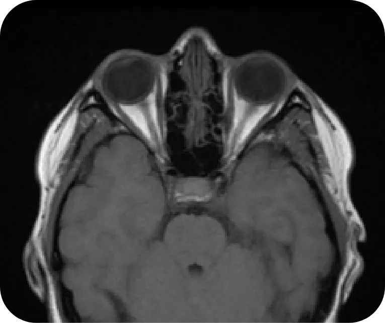
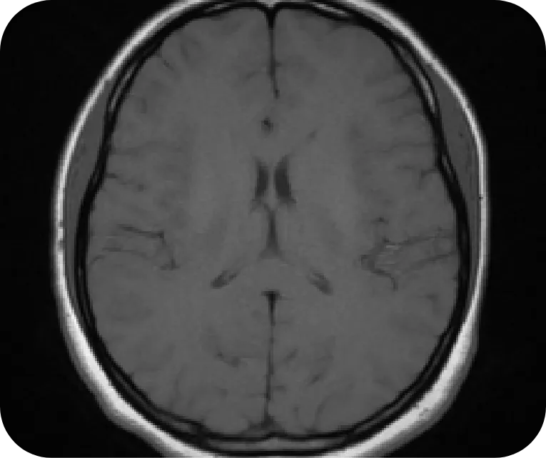
Gaillard F. Normal brain (MRI). Radiopaedia. Published July 2, 2015. Accessed July 26, 2022. https://radiopaedia.org/cases/normal-brain-mri-6?lang=us 14. 
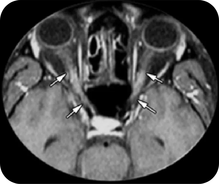
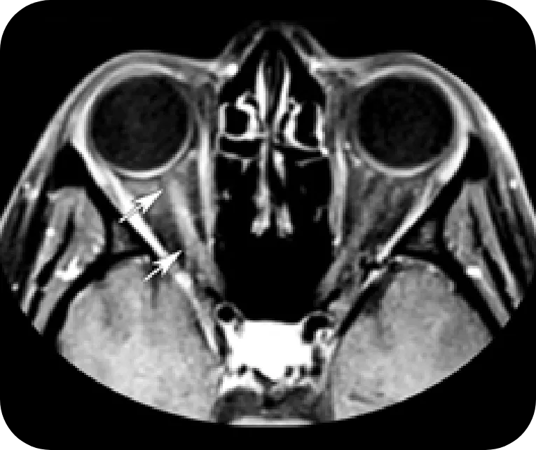
Dutra BG, da Rocha AJ, Nunes RH, Maia ACM Jr. Neuromyelitis optica spectrum disorders: spectrum of MR imaging findings and their differential diagnosis. Radiographics. 2018;38(1):169-193. 15. 
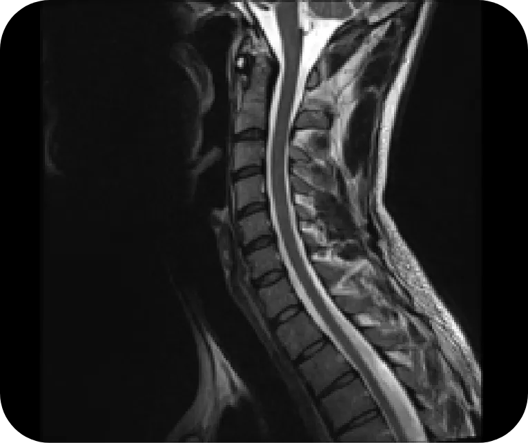
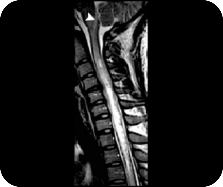
Di Muzio B. Normal cervical spine MRI. Radiopaedia. Published July 19, 2015. Accessed July 26, 2022. https://radiopaedia.org/cases/normal-cervical-spine-mri-1?lang 16. Trebst C, Jarius S, Berthele A, et al. Update on the diagnosis and treatment of neuromyelitis optica: recommendations of the Neuromyelitis Optica Study Group (NEMOS). J Neurol. 2014;261(1):1-16. 17. Kitley J, Leite MI, Nakashima I, et al. Prognostic factors and disease course in aquaporin-4 antibody-positive patients with neuromyelitis optica spectrum disorder from the United Kingdom and Japan. Brain. 2012;135(pt 6):1834-1849.